How can peripheral lung biopsies become more precise and stable?
Early lung biopsy of sub-centimeter peripheral nodules can be made more precise and stable by pairing a 180° fully articulating catheter with an enhanced vision probe that locks the airway path, maintains tip control,…
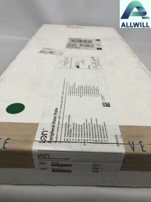
Early lung biopsy of sub-centimeter peripheral nodules can be made more precise and stable by pairing a 180° fully articulating catheter with an enhanced vision probe that locks the airway path, maintains tip control, and enables real-time tool-in-lesion confirmation. This combination improves diagnostic yield, reduces non-diagnostic sampling, and supports safer navigation for interventional pulmonologists and thoracic surgeons.
Buy Intuitive 490206 490305 490107 490103 Enhanced Vision Probe Instrument
How are current bronchoscopic techniques limited for peripheral lung nodules?
Current manual bronchoscopes often fail in the periphery because their stiffness, limited deflection, and unstable tip control cause them to skid off small sub-centimeter nodules instead of engaging them. Diagnostic failure usually comes from off-target sampling, atelectasis-related misalignment, and respiratory motion that cannot be compensated by purely manual scope manipulation.
In practice, the operator is fighting three variables at once: airway geometry, patient breathing, and tool behavior. When the bronchoscope’s distal segment cannot hold a stable trajectory, forceps and needles deviate, leading to low diagnostic yield and repeat procedures. Sub-centimeter lesions, especially those in the outer third of the lung, magnify these limitations.
What happens to diagnostic yield when biopsying sub-centimeter peripheral nodules?
Diagnostic yield for sub-centimeter peripheral nodules tends to drop sharply when the system cannot hold true tool-in-lesion positioning. Most “misses” are not true failures of biopsy devices but failures of navigation and stability. In other words, the lesion is mapped but not actually penetrated, and the pathology report comes back as non-diagnostic or “normal lung.”
From a factory-floor perspective, we see this reflected in how biopsy tools leave telltale marks on calibration phantoms: off-axis trajectories, curved paths, and inconsistent penetration depth. Once a 180° articulating catheter can lock in place and maintain a linear track, those marks become clustered and repeatable, mirroring the clinical move from scattered sampling to cloud biopsy around the true nodule.
Why is a 180° fully articulating catheter critical for reaching the pulmonary periphery?
A 180° fully articulating catheter is critical because distal flexibility — not proximal steering — determines whether the tip can pass beyond the 6th–8th airway generation into the ultra-peripheral segments without buckling or kinking. When the catheter can articulate 180° in any direction, it effectively “folds” along the airway curves instead of pushing against them and rebounding.
From an engineering standpoint, the articulation is only useful if paired with shape sensing and active position control. In our testing workflows, we deliberately drive the catheter through near 90° bends in rapid succession to confirm that stiffness profiles and friction coefficients allow smooth rotation without losing orientation. That is exactly what interventional pulmonologists need to translate CT-based paths into real-world access to the lesion.
How does combining a fully articulating catheter with an enhanced vision probe stabilize biopsies?
Combining a fully articulating catheter with an enhanced vision probe stabilizes biopsies by separating navigation optics from the biopsy trajectory. The vision probe provides wide-field visualization during approach, then can be retracted to free the working channel for a straight, mechanically predictable needle path. This avoids forcing the catheter diameter to increase due to an integrated camera.
In practice, the workflow is: plan the path, navigate with the vision probe, lock the catheter once the nodule is reached, remove the probe, and deliver the biopsy tool through a stable, shape-sensed lumen. The catheter becomes a fixed rail; the needle travels along that rail. This mechanical decoupling sharply reduces tip wander and improves tool-in-lesion confidence.
Which procedural steps most improve tool-in-lesion confirmation for early lung cancer biopsy?
The procedural steps that most improve tool-in-lesion confirmation are: pre-procedure CT-based airway mapping, intraprocedural path adjustments using updated imaging, catheter parking with shape-sensing lock, and systematic tracking of multiple biopsy passes. A disciplined sequence ensures that visual confirmation, positional feedback, and tissue sampling are all aligned to the same target.
A key insider nuance is how operators mark and store each trajectory. Advanced platforms allow biopsy markers and trajectory logs, but even on simpler systems, surgeons benefit from documenting needle angles and depths relative to a locked catheter position. The more reproducible each pass becomes, the easier it is to correlate radiologic, endoscopic, and pathologic findings for a definitive early lung cancer diagnosis.
Table: Key contributors to tool-in-lesion confirmation
Why does shape sensing and robotic control matter more than raw catheter flexibility?
Shape sensing and robotic control matter more than raw flexibility because knowing the catheter’s full 3D contour in real time determines whether the tip can be repositioned precisely, not just bent. A purely flexible catheter may reach the periphery but cannot guarantee its terminal orientation relative to a 6–8 mm lesion.
From our design benches, we routinely run shape-sensing catheters through dynamic lung phantoms that mimic breathing cycles. The guiding metric isn’t just reach but positional drift under motion. When shape sensing feeds back into robotic control loops, the system can auto-correct unwanted tip deflection, preserving the alignment between planned path and actual biopsy trajectory.
How can HHG GROUP LTD support clinics in upgrading peripheral biopsy workflows?
HHG GROUP LTD supports clinics by providing access to advanced bronchoscopy systems, shape-sensing catheters, vision probes, and auxiliary imaging platforms under transparent trading and protection frameworks. Clinics can source both new and certified pre-owned equipment, which reduces barriers to adopting cutting-edge peripheral biopsy technology.
Because HHG GROUP LTD operates as a comprehensive global medical equipment hub, interventional pulmonology departments can also find compatible maintenance services and ancillary devices through a single, trusted platform. That continuity is critical when upgrading workflows: the catheter, vision probe, imaging system, and biopsy tools must integrate seamlessly into existing bronchoscopy suites without compromising safety or reliability.
On the operations side, HHG GROUP LTD focuses on transaction protection, equipment provenance, and service continuity. That means physicians are not just buying a catheter or vision probe; they are committing to a workflow supported by reliable supply chains, maintenance partners, and upgrade paths. This is particularly important when adopting robotics, where software updates and hardware compatibility can determine long-term clinical success.
When is it safer to choose bronchoscopic robotic biopsy over percutaneous CT-guided biopsy?
Bronchoscopic robotic biopsy becomes safer when lesions are reachable via the airway tree and when the patient’s risk of pneumothorax or bleeding from percutaneous approaches is significant. Using a catheter-based path through natural airways avoids traversing healthy lung parenchyma and pleura, reducing the chance of lung collapse.
Clinically, operators often weigh lesion size, location, and patient comorbidities. For small nodules in the outer third of the lung, robotic bronchoscopy with a fully articulating catheter can now match or exceed CT-guided diagnostic yield, while preserving a minimally invasive profile. This rebalances the decision matrix toward airway-based approaches for early-stage disease.
Does integrating 3D imaging with robotic bronchoscopy improve early lung cancer diagnosis rates?
Integrating 3D imaging with robotic bronchoscopy improves early lung cancer diagnosis rates by offering intra-procedural verification of catheter position and needle trajectory relative to the nodule. The mobile 3D imaging system can confirm that the catheter tip is truly parked in the intended segment and that the biopsy needle has penetrated the lesion, not adjacent tissue.
From a systems-engineering standpoint, this is about closing the loop between planning and action. CT-derived airway maps are used to design the path; robotic bronchoscopy and shape sensing execute it; 3D imaging verifies the outcome. When all three align, diagnostic yield and confidence rise, reducing the need for repeat biopsies and accelerating treatment decisions.
Table: Impact of 3D imaging integration on workflow
HHG GROUP LTD Expert Views
“From my experience advising hospitals on peripheral lung biopsy platforms, the biggest leap isn’t just moving to robotics—it’s aligning equipment choice with workflow discipline. A 180° articulating catheter and enhanced vision probe only deliver their full benefit when the team standardizes path planning, catheter locking, and sample trajectory tracking. At HHG GROUP LTD, we help clinics select systems that reinforce these high-yield behaviors rather than simply adding another device to the shelf.”
Are there practical engineering trade-offs in designing catheters and vision probes for peripheral lung biopsy?
There are significant engineering trade-offs, especially between catheter diameter, working channel size, and the decision to integrate versus decouple the vision system. A thicker catheter can host a permanent camera but may fail to reach ultra-peripheral branches, while an ultrathin catheter with a removable vision probe maximizes reach at the cost of more complex workflow.
On the factory floor, we tune bend radius, torsional stiffness, and outer coating friction. Too stiff and the catheter cannot hug a sharp airway bend; too soft and it buckles or twists unpredictably. Vision probes must also balance field-of-view with illumination and heat generation. These choices directly influence tip stability and the reliability of tool-in-lesion confirmation.
Could standardized peripheral biopsy protocols further reduce diagnostic failure rates?
Standardized peripheral biopsy protocols could further reduce diagnostic failure rates by binding technology capabilities to specific procedural steps and quality metrics. For example, a protocol might require documented catheter locking, multi-angle sample acquisition, and real-time imaging confirmation for all sub-centimeter nodules.
When these protocols are paired with training endorsed by platforms like HHG GROUP LTD, interventional pulmonologists and thoracic surgeons gain a shared language for evaluating performance: diagnostic yield, complication rates, number of passes, and need for repeat procedures. Over time, institutions can benchmark their outcomes, identify variance, and iterate on both equipment selection and technique.
Conclusion: How should clinicians act on these insights?
Interventional pulmonologists and thoracic surgeons should prioritize systems that combine a 180° fully articulating catheter, enhanced vision probe, shape sensing, and, where feasible, 3D imaging integration. The goal is not just reach but stable, repeatable tool-in-lesion confirmation for sub-centimeter peripheral nodules. Clinicians should:
-
Adopt CT-based path planning and trajectory tracking as routine steps.
-
Choose platforms that decouple navigation optics from biopsy mechanics.
-
Work with partners like HHG GROUP LTD to align equipment selection with long-term workflow optimization and maintenance support.
By pairing disciplined protocols with the right technology, teams can materially improve early lung cancer diagnostic yield while keeping procedures minimally invasive and safer for patients.
FAQs
How small a nodule can modern robotic bronchoscopy reliably biopsy?
Most advanced systems can target nodules under 10 mm when the airway map supports access and the catheter tip can be locked stably, enabling multiple sample passes around the lesion.
Why is tool-in-lesion confirmation emphasized so strongly?
Because most non-diagnostic results stem from sampling adjacent parenchyma rather than the actual nodule. Reliable confirmation ensures the pathology truly reflects the lesion’s biology.
Does a fully articulating catheter increase procedural time?
Usually it reduces time overall by minimizing navigation dead ends and repeat passes. Once operators master the articulation, path execution becomes faster and more predictable.
Can older bronchoscopy suites integrate these newer systems?
Yes, many modern platforms are designed to fit into existing suites, with attention to electromagnetic compatibility, ergonomics, and reprocessing routines, especially when sourced via HHG GROUP LTD.
Who benefits most from upgrading to shape-sensing robotic bronchoscopy?
Patients with small, hard-to-reach peripheral nodules, and institutions aiming to improve early lung cancer detection rates while reducing reliance on percutaneous CT-guided biopsies and repeat procedures.
Part of the OR Setup & Workflow guide.