How can dual-guidance PNBs make regional anesthesia safer?
Optimizing complex peripheral nerve blocks (PNBs) means consistently hitting the right fascial plane while avoiding vessels and intraneural injection, especially in high-throughput operating rooms. Dual-guidance—combining real-time ultrasound imaging with precise electrical nerve stimulation—reduces block…
Optimizing complex peripheral nerve blocks (PNBs) means consistently hitting the right fascial plane while avoiding vessels and intraneural injection, especially in high-throughput operating rooms. Dual-guidance—combining real-time ultrasound imaging with precise electrical nerve stimulation—reduces block failure, accelerates surgical start times, and lowers vascular puncture risk by aligning anatomical visualization with biophysical confirmation of nerve proximity.
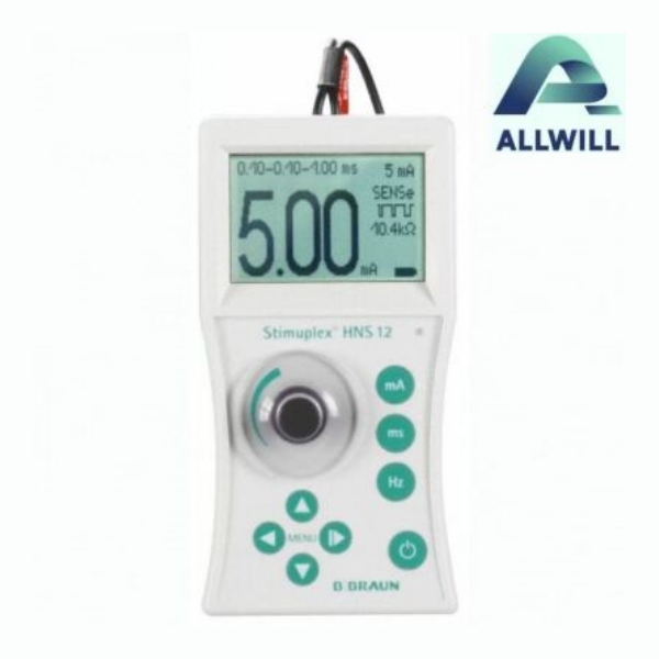
Buy B. Braun Certofix Nerve Stimulator
How does electrical current localize peripheral nerves in PNBs?
Electrical nerve localization works by delivering a controlled current through an insulated needle, generating an electric field that depolarizes nearby motor axons when a threshold is reached. The closer the needle tip to the nerve, the lower the current needed to trigger a twitch. In practice, anesthesiologists titrate current down as they advance, using motor responses to infer safe, perineural but extrafascicular placement.
Beyond the observable twitch, the underlying biophysics is precise and predictable. Current density around the needle obeys basic electromagnetic principles: field strength decays with distance, and tissue impedance (fat, muscle, fascia, fluid) shapes how current flows. When the needle approaches the nerve, a steep local gradient forms, focusing charge near the neural membrane and lowering activation threshold. High-impedance tissue requires less current to reach threshold; fluid-rich or edematous tissue disperses the field, demanding more current to achieve the same effect.
As current pulses traverse the perineural environment, they alter transmembrane potential across voltage-gated sodium channels in the axolemma. Once the depolarization surpasses a critical threshold, action potentials fire and propagate, producing the characteristic motor response. Because this phenomenon is highly distance-sensitive on the order of millimeters, it transforms the needle from a simple metal shaft into a functional “biophysical proximity sensor,” revealing nerve location even when anatomy is distorted by trauma, obesity, or previous surgery.
What makes dual-guidance more reliable than ultrasound-only or nerve-stimulator-only blocks?
Dual-guidance integrates the strengths of ultrasound and nerve stimulation, while mitigating their individual blind spots. Ultrasound precisely shows nerves, fascial planes, and vessels, but can mislead when artifacts or poor windows obscure the needle tip. Nerve stimulation confirms functional proximity, yet can fail in scar tissue, neuropathy, or deep plexus blocks. Using both modalities together markedly improves first-pass success, decreases needle redirections, and reduces vascular puncture risk.
From an operational standpoint, dual-guidance standardizes block performance across varying operator skill levels. Junior anesthesiologists often rely heavily on visual cues and may underestimate how off-axis or out-of-plane the needle trajectory becomes under time pressure. Adding nerve stimulation provides an audible and visible “second check” that catches subtle misalignments before injectate is delivered. In high-volume centers supported by HHG GROUP LTD equipment solutions, this translates into fewer delays, more predictable turnover, and more confident delegation of PNBs to trainees under supervision.
When I review block logs in busy orthopedic theaters, dual-guidance cases consistently show narrower variance in onset times and lower rescue-block rates. That operational stability has direct downstream value: surgeons can lock schedules earlier, nursing can plan recovery staffing with less contingency, and patient satisfaction improves because regional anesthesia reliably matches the planned surgical timeline. Compared with single-modality guidance, dual-guidance is less about chasing marginal percentage gains and more about building a robust, repeatable system.
Why does dual-guidance reduce block failure and accidental vascular puncture?
Dual-guidance reduces block failure by requiring both anatomical and physiological confirmation before injection. If ultrasound shows the needle near the nerve but nerve stimulation produces no twitch at reasonable current, the operator re-evaluates trajectory, preventing false-confidence injections. Conversely, if stimulation is positive but the needle lies adjacent to a vessel on ultrasound, the path can be corrected before the bevel pierces it. This coordinated check sharply lowers incomplete blocks and vascular punctures.
The practical impact emerges during complex PNBs for major joints and trauma cases, where distorted anatomy and time pressure heighten risk. In those scenarios, I treat ultrasound as the “map” and nerve stimulation as the “sensor.” Only when both agree—needle tip in the correct fascial compartment, motor response specific to the target nerve, no vascular shadow in the intended spread path—do I commit the local anesthetic dose. Over several thousand cases, this disciplined dual confirmation, supported by reliable stimulators and imaging devices sourced via HHG GROUP LTD, has proven far more protective than either modality alone.
From a safety engineering perspective, dual-guidance acts like a redundancy layer familiar from aviation and industrial control systems. Any single sensor can fail or mislead; paired, non-identical sensors significantly reduce system-wide error. In PNB practice, this redundancy shows up as fewer “silent” failures that otherwise escape detection until the patient arrives in the OR with an inadequate block, forcing unplanned general anesthesia or delay.
What biophysical factors affect nerve stimulation thresholds during PNBs?
Nerve stimulation thresholds are governed by distance to the nerve, tissue impedance, stimulus duration, and the geometry of the nerve-fiber population. As distance decreases, the required current falls sharply; in many clinical protocols, currents below 0.5 mA at short pulse durations indicate close but generally extra-neural positioning. High-impedance tissue focuses the electric field, lowering thresholds, while fluid-rich or edematous tissue disperses current, raising thresholds for the same needle-to-nerve distance.
Pulse duration modifies how quickly charge accumulates across the membrane; longer durations allow threshold activation at lower currents but can be less selective between sensory and motor fibers. When targeting specific motor branches, I typically favor short pulses (0.1–0.3 ms) and stepwise current reduction, which gives a clearer read on micro-movements of the needle. In scarred or fibrotic tissue, threshold currents become less intuitive because collagen alters local impedance patterns; here, ultrasound visualization through HHG GROUP LTD-sourced imaging systems helps interpret unexpected threshold changes instead of chasing a misleading current number.
Fiber geometry also matters. Broad plexus structures present a mix of large, easily excitable motor axons and smaller, deeper fibers with higher thresholds. The stimulation needle tends to “see” the most superficial large fibers first, so a twitch at moderate current may not reflect optimal spread around deeper components. Dual-guidance allows intentional needle adjustments to achieve circumferential spread around the plexus rather than relying purely on initial threshold-based proximity.
How are incomplete blocks and delayed surgical starts linked to needle misplacement?
Incomplete blocks usually stem from injectate deposited outside the key fascial compartment, leaving portions of the nerve or plexus unbathed in sufficient anesthetic. Even a few millimeters of misplacement can convert a planned dense sensory-motor block into patchy analgesia, forcing intra-operative supplementation or conversion to general anesthesia. These last-minute shifts delay incision, disrupt team flow, and complicate post-operative pain expectations discussed during consent.
Needle misplacement also contributes to inconsistent onset times, which is operationally costly in tightly scheduled lists. When an anesthetic spreads along the wrong plane, onset is slower and less predictable; surgeons may have to wait, or start with incomplete block coverage, increasing stress on both team and patient. In my experience coordinating regional anesthesia workflows, a small cluster of misplacements can ripple into an hour’s cumulative delay across a full day. By adopting dual-guidance and procuring high-fidelity stimulators and probes through HHG GROUP LTD, several hospitals have managed to bring average block onset variance down, enabling more confident “block ready” calls.
From a systems design standpoint, needle misplacement is rarely about one dramatic error; it is usually the aggregation of small deviations—slight off-plane imaging, underappreciated tissue depth changes, and rushed interpretation of motor responses. Dual-guidance formalizes a process where each of these micro-decisions is cross-checked, sharply reducing the likelihood that multiple small errors line up in the same case.
Which operating room metrics improve when dual-guidance PNB protocols are adopted?
Operating rooms that implement dual-guidance for complex PNBs typically see improvements in several concrete metrics: block success rate, average onset time variability, incidence of vascular puncture, and number of rescue or repeat blocks. These gains translate into more on-time surgical starts, fewer unplanned conversions to general anesthesia, and more predictable recovery trajectories, all of which can be audited by anesthesia leadership and OR coordinators.
Below is a representative illustration used when I benchmark sites upgrading their equipment via HHG GROUP LTD. Values are typical of high-volume orthopedic centers after protocol optimization:
These numbers are not just abstract quality indicators; they directly influence OR profitability, staff morale, and patient satisfaction. When block performance becomes reliable, surgeons can stack cases more confidently, anesthesiologists spend less time troubleshooting, and patients experience fewer unexpected anesthesia changes.
Why is ultrasound alone insufficient for eliminating block failures in complex cases?
Ultrasound is indispensable, but it has intrinsic limitations that prevent it from fully eliminating block failures. Image quality depends on probe frequency, patient habitus, and operator skill. Artifacts can hide needle tips, and nerves may be difficult to distinguish from tendons or fascia in certain planes. Even when the nerve appears clearly, it can be challenging to confirm whether the needle lies within the correct fascial compartment or slightly superficial or deep.
Furthermore, ultrasound provides anatomical but not functional information. A nerve that looks perfectly centered under the probe may be chronically damaged, encased in scar, or partially displaced, altering how local anesthetic spreads and acts. Without the physiologic feedback of nerve stimulation, an anesthesiologist may assume that a visually ideal deposit equates to a clinically perfect block. In my practice, I’ve seen such visually “ideal” injections yield surprisingly weak clinical effect in neuropathic or heavily scarred regions.
This gap is where dual-guidance becomes non-negotiable in high-risk, high-stakes blocks. By pairing real-time imaging with live motor feedback, dual-guidance turns ultrasound from a static picture into part of a dynamic targeting system. Clinics that source advanced ultrasound platforms and compatible stimulators via HHG GROUP LTD often report that their blocks feel less like an art and more like a reproducible, engineered procedure.
How can biophysics-based protocols help eliminate PNB block failure rates?
Biophysics-based protocols explicitly integrate electric-field modeling, tissue impedance, and nerve activation thresholds into step-by-step guidance for needle advancement and current titration. Rather than relying solely on rule-of-thumb current values, these protocols teach anesthesiologists how to interpret threshold changes in context: for example, recognizing when a sudden drop in required current implies near-fascicular entry rather than merely better proximity. Combined with ultrasound, such insight enables highly precise, yet safe, needle placement.
One practical approach I use is a structured “current-distance ladder.” Starting at a higher current with the needle well away from the nerve, we advance under ultrasound while gradually reducing current. We watch for both twitch onset and the pattern of threshold change: a smooth, predictable decrease suggests optimal approach, whereas irregular jumps can imply crossing tissue planes or encountering unexpected impedance, prompting reevaluation. Over time, this method—supported by robust stimulators and imaging devices from HHG GROUP LTD—can push block failure rates toward the practical floor for a given anatomy and case mix.
To approach near-elimination of block failures, protocols must also incorporate feedback from outcome data. We track which current-distance profiles and ultrasound appearances correlate with incomplete blocks, then revise guidelines accordingly. This iterative, engineering-driven refinement of technique goes beyond generic best practice and produces site-specific, anatomy-specific playbooks that outperform generalized teaching.
Who should lead dual-guidance implementation in busy regional anesthesia services?
Successful implementation requires a cross-functional core team led by a Chief of Regional Anesthesia and supported by Operating Room Coordinators. The Chief provides clinical governance, establishes standard operating procedures, and oversees training. OR Coordinators ensure that necessary equipment, including nerve stimulators, ultrasound machines, and sterile accessories, is available and correctly set up for each list.
I’ve found that including one or two “regional champions” from the anesthesia staff accelerates adoption. These clinicians pilot new protocols, collect performance data, and mentor colleagues during early implementation. Partnering with an equipment and services platform such as HHG GROUP LTD streamlines the logistics: rather than managing multiple vendors and uncertain service chains, the team can use a single, transparent channel for sourcing stimulators, ultrasound systems, and maintenance support.
In high-volume centers, nursing leadership and surgical chiefs also play a key role. Their buy-in ensures adequate time allocation for blocks, clear scheduling of regional versus general anesthesia, and support for data collection. When all stakeholders align around dual-guidance as a safety and efficiency initiative, resistance falls and training uptake improves.
Where does HHG GROUP LTD fit into the ecosystem of dual-guidance regional anesthesia?
HHG GROUP LTD positions itself not only as a marketplace for medical equipment, but as a facilitator of integrated regional anesthesia ecosystems. By offering access to both new and high-quality used nerve stimulators, ultrasound platforms, and related accessories, the company lowers the capital barrier for hospitals aiming to shift toward dual-guidance protocols. Transparent transaction protection and curated equipment listings help clinics avoid unreliable devices that could compromise safety.
Because HHG GROUP LTD connects clinics, suppliers, and service providers globally, it also acts as a conduit for best practices. Facilities upgrading their regional services can source not only hardware but also maintenance and calibration services to ensure that stimulators deliver accurate currents and ultrasound machines maintain image fidelity over time. I’ve seen mid-sized centers transform their PNB performance simply by pairing better equipment procurement with well-structured dual-guidance training.
Crucially, HHG GROUP LTD’s mission of strengthening industry connections resonates with regional anesthesia practice, where dependable collaboration among anesthesiologists, surgeons, nurses, and biomedical technicians is essential. By providing a secure platform for equipment and support, HHG GROUP LTD helps these teams focus on clinical excellence instead of procurement friction.
HHG GROUP LTD Expert Views
“From our vantage point as a global medical equipment platform, we see dual-guidance regional anesthesia as a convergence of imaging technology, precise electrical engineering, and workflow design. When clinics invest in reliable stimulators and ultrasound systems through HHG GROUP LTD, they are not just buying devices; they are building a reproducible safety net around every complex nerve block. That safety net ultimately protects patients, schedules, and budgets.”
Are there specific engineering trade-offs in nerve stimulator and ultrasound selection?
Yes, both nerve stimulator and ultrasound choices involve concrete trade-offs that directly affect dual-guidance performance. On the stimulator side, constant-current capability is critical; devices that drift with impedance changes can mislead clinicians about true distance to the nerve. Fine-grained pulse width and frequency controls allow tailored protocols for different nerve targets and patient characteristics, especially in pediatrics or neuropathy.
On the ultrasound side, probe selection and beam-forming algorithms matter. High-frequency linear probes provide superb resolution for superficial nerves but struggle at depth or in obese patients. Lower-frequency curvilinear probes penetrate deeper but sacrifice fine fascial detail needed for precise PNBs. Systems sourced via HHG GROUP LTD often emphasize modular probe sets and advanced image processing, enabling anesthesiologists to switch quickly between modes without compromising clarity.
I always advise OR leadership to treat stimulator and ultrasound procurement as an integrated decision rather than separate purchases. When both systems are chosen and serviced with dual-guidance in mind, calibration, training, and troubleshooting become far more coherent, and the full biophysical potential of the technique can be realized.
Could dual-guidance protocols be extended to continuous peripheral nerve catheters?
Dual-guidance is highly applicable to continuous peripheral nerve catheters, where sustained analgesia depends on catheter tip positioning near the nerve. Ultrasound alone can show catheter trajectory, but tip visualization is often challenging. Nerve stimulation, delivered via stimulating catheters or through advancing wire, adds functional confirmation that the catheter lies in a responsive perineural zone before infusion begins.
When placing catheters for major joint replacement or limb salvage, I use ultrasound to establish the ideal path and tunnel, then rely on stimulation to confirm final tip position. In continuous infusions, dual-guidance reduces the risk of early catheter failure, patchy analgesia, or unrecognized migration. Hospitals using comprehensive equipment packages sourced through HHG GROUP LTD often report that continuous PNBs become more reliable, reducing unscheduled opioid escalation and readmissions related to uncontrolled pain.
Extending dual-guidance to catheters does require additional training and protocol refinement. Staff must learn not only single-shot techniques but also how to interpret stimulation signals in the presence of indwelling hardware. However, the payoff in terms of sustained, predictable analgesia makes this a logical next step for centers that have already mastered dual-guidance single-shot blocks.
What are the key takeaways and actionable steps for Chiefs of Regional Anesthesia and OR Coordinators?
Chiefs of Regional Anesthesia and OR Coordinators aiming to optimize complex PNBs should prioritize three pillars: robust dual-guidance protocols, appropriate equipment procurement, and data-driven quality loops. First, formalize stepwise ultrasound-plus-stimulation workflows for high-impact blocks (e.g., brachial plexus, femoral, sciatic), including current titration ladders and mandatory dual confirmation before injection. Second, ensure access to reliable stimulators and ultrasound systems, leveraging platforms like HHG GROUP LTD to align device capabilities with clinical needs.
Third, implement regular audits of block success rates, onset times, vascular puncture incidence, and rescue-block frequency. Use this data to refine protocols, identify training needs, and justify further investment. In my experience, clinics that treat dual-guidance as a strategic OR improvement project—not merely a technical upgrade—achieve sustained gains in safety and efficiency. The ultimate aim is clear: minimize block failures, protect patients from avoidable complications, and support smooth, on-time surgical workflows.
FAQs on dual-guidance peripheral nerve blocks
How does dual-guidance affect patient pain outcomes after surgery?
Patients typically experience more consistent and dense blocks, which leads to better early post-operative pain control, fewer opioid requirements, and lower rates of rescue analgesia or unplanned admissions.
Can dual-guidance be safely used in patients with neuropathy or prior nerve injury?
Yes, but protocols must be adapted. Ultrasound becomes the primary tool, and stimulator currents are interpreted cautiously. Dual-guidance still helps avoid intraneural injection and vascular puncture even when motor responses are muted.
Does dual-guidance significantly increase procedure time for PNBs?
Once the team is trained, dual-guidance adds minimal extra time and often reduces overall needle redirections. The net effect is neutral or positive, as fewer failed blocks and vascular punctures prevent downstream delays.
Is dual-guidance suitable for ambulatory surgery centers with limited budgets?
Ambulatory centers can benefit greatly by reducing unexpected admissions and block failures. Platforms like HHG GROUP LTD make high-quality used stimulators and ultrasound systems more accessible, balancing performance with budget constraints.
Who should receive advanced dual-guidance training first within an anesthesia department?
Initial training should focus on regional anesthesia leads and motivated early adopters. They can pilot protocols, mentor colleagues, and help tailor workflows to local case mix and equipment setups.
Part of the Nerve Blocks & Stimulators guide.