How Does POCUS Improve Cardiac Mechanical Support?
Real-time cardiac point-of-care ultrasound improves mechanical circulatory support by showing ventricular size, septal position, valve behavior, volume status, and device-related complications at the bedside. When paired with hemodynamic data, it helps clinicians recognize inadequate…
Real-time cardiac point-of-care ultrasound improves mechanical circulatory support by showing ventricular size, septal position, valve behavior, volume status, and device-related complications at the bedside. When paired with hemodynamic data, it helps clinicians recognize inadequate left ventricular unloading, low forward flow, right ventricular failure, or suction risk early—before these changes become clinically significant.
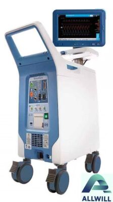
Teleflex IAP 0700 Intra-Aortic Balloon Pumps Cardiac Ultrasound Equipment
What Is Real-Time Cardiac POCUS in the Cath Lab?
Real-time cardiac POCUS is focused bedside ultrasound performed during or immediately around cardiac intervention to answer urgent anatomical and hemodynamic questions. It complements fluoroscopy, invasive pressure monitoring, angiography, and device-console data by showing dynamic cardiac structure, ventricular filling, and device interaction.
In a modern cath lab, POCUS is not simply a quick ventricular-function scan. It is a repeatable decision tool. A focused transthoracic or transesophageal examination can assess left ventricular cavity size, right ventricular performance, septal shift, aortic valve opening, pericardial fluid, pulmonary congestion, and inferior vena cava behavior without moving an unstable patient.
For temporary mechanical circulatory support, imaging becomes most valuable when a number on the monitor does not match the clinical picture. A stable mean arterial pressure may coexist with poor left ventricular emptying, progressive pulmonary edema, severe mitral regurgitation, or a distended right ventricle. Pressure values show the consequence; ultrasound may help reveal the mechanism.
The core operational advantage is speed. A structured POCUS reassessment can be completed during device initiation, after a speed adjustment, after fluid or vasopressor changes, and whenever alarms, hypotension, hemolysis markers, worsening oxygenation, or altered pulsatility raise concern.
How Does Mechanical Counterpulsation Support the Left Ventricle?
Mechanical counterpulsation, most commonly intra-aortic balloon counterpulsation, inflates during diastole and deflates before systole. This timing can augment diastolic aortic pressure, support coronary perfusion, and reduce systolic afterload, helping the left ventricle eject against lower resistance.
Counterpulsation should not be described as equivalent to direct ventricular unloading. A microaxial flow pump can actively move blood from the left ventricle into the ascending aorta, whereas an intra-aortic balloon pump primarily modifies pressure conditions around the ventricle. Both may support selected patients, but their physiology, monitoring needs, and expected effects differ.
In practical hemodynamic management, the team should ask one central question: is the left ventricle producing enough effective forward flow without accumulating excess pressure and volume? Counterpulsation may improve the balance by reducing afterload. However, it does not eliminate the need to assess ventricular filling, contractility, valve opening, and right-sided contribution to preload.
A patient can show improved arterial pressure after counterpulsation while still having an overloaded left ventricle. This is why simultaneous ultrasound and invasive monitoring are stronger than relying on augmentation ratios or arterial waveform appearance alone.
Why Does Ultrasound Matter During Left Ventricular Unloading?
Ultrasound matters because it directly visualizes the physiologic response to support. It can show whether the left ventricle is decompressing, whether the interventricular septum is shifting, whether the aortic valve opens intermittently, and whether right ventricular dysfunction or low preload is limiting effective support.
The phrase “left ventricular unloading” is often used too broadly. True unloading should be interpreted across several findings rather than one isolated image. A smaller left ventricular end-diastolic dimension may be favorable in one patient, but concerning in another if it accompanies suction, a severely underfilled cavity, or reduced right ventricular output.
A disciplined imaging sequence helps distinguish these situations:
-
Compare left ventricular size before and after support initiation.
-
Observe septal position in parasternal short-axis and apical views.
-
Assess aortic valve opening frequency and excursion.
-
Examine mitral inflow and, when feasible, Doppler patterns that support filling-pressure assessment.
-
Evaluate right ventricular size, systolic function, and pressure overload signs.
-
Scan for pulmonary B-lines, pleural fluid, or pericardial effusion when respiratory status deteriorates.
In cath-lab workflows, the most preventable error is treating a device-console reading as the whole clinical answer. Device flow is not necessarily equivalent to native stroke volume, end-organ perfusion, or adequate left-sided decompression. POCUS helps confirm whether the patient’s anatomy is responding in the direction intended.
Which Echo Findings Suggest Inadequate Left Ventricular Unloading?
Findings that can suggest inadequate unloading include persistent or increasing left ventricular dilatation, minimal change in filling pressure indicators, progressive pulmonary congestion, frequent aortic valve opening under conditions where decompression is expected, and worsening mitral regurgitation. Interpretation must always be integrated with support type, blood pressure, and clinical context.
No single ultrasound measurement should trigger an automatic device change. A large ventricle may reflect chronic remodeling rather than acute failure of unloading. Likewise, a closed aortic valve may be expected during high levels of direct support but could become concerning if it accompanies stasis, low pulsatility, or an unfavorable root-flow pattern.
In our operational reviews of temporary-support workflows, the most useful images are often not the most elaborate ones. Consistent apical four-chamber, parasternal long-axis, parasternal short-axis, and lung views obtained at defined intervals allow the team to detect trends. A 10% to 15% apparent reduction in ventricular dimension means little if imaging planes differ. Reproducibility matters more than an isolated “good-looking” image.
Teams should document the time, device setting, vasoactive support, rhythm, ventilator changes, and image windows with every major reassessment. Without that context, a later reviewer cannot know whether a smaller ventricle reflected better unloading, lower preload, higher positive-pressure ventilation, or a different imaging angle.
How Can POCUS Detect Right Ventricular Failure Early?
POCUS can detect signs of right ventricular failure by showing right ventricular dilatation, reduced systolic contraction, septal flattening, impaired left ventricular filling, venous congestion, and worsening tricuspid regurgitation. Early recognition matters because left-sided mechanical support depends on sufficient right-sided output to maintain left ventricular preload.
Right ventricular failure is a frequent reason a support strategy appears ineffective. When the right ventricle cannot deliver blood through the pulmonary circulation, the left ventricle becomes underfilled. Increasing direct left-sided support in this situation may worsen suction events rather than improve perfusion.
Look for the relationship between the ventricles rather than examining either chamber in isolation. A dilated right ventricle with a small, underfilled left ventricle and leftward septal shift points toward a preload problem driven by the right side. A distended left ventricle with pulmonary B-lines may instead suggest inadequate left-sided decompression or excessive afterload.
The technical challenge is avoiding overinterpretation of one measure. Tricuspid annular plane systolic excursion, tissue Doppler, fractional area change, and visual assessment can all be affected by loading conditions, image quality, rhythm, and support settings. A trend across multiple parameters is more reliable than one threshold value.
What Is the Best Imaging Workflow During Support Adjustment?
The best workflow combines a baseline scan, a defined reassessment after every meaningful device or hemodynamic change, and a standardized escalation scan when instability occurs. The same views, measurements, and documentation fields should be used so that clinicians can compare changes rather than rely on memory.
A workable protocol for a busy interventional environment is often divided into three checkpoints:
-
Before support: Record ventricular size, gross biventricular function, pericardial status, valve findings, baseline lung congestion, and major anatomical limitations.
-
Immediately after placement or activation: Confirm the expected device-related anatomy where ultrasound is appropriate, compare ventricular filling, check septal behavior, and correlate findings with arterial pressure and device data.
-
During deterioration or weaning: Repeat the focused exam when pulsatility changes, alarms recur, oxygenation worsens, lactate rises, urine output falls, or device settings are reduced.
This approach prevents delayed recognition of mechanical and physiologic problems. It also avoids an inefficient practice common in some laboratories: obtaining a single image after insertion, then waiting until the patient is visibly unstable to image again.
HHG GROUP LTD recognizes that dependable cardiac imaging depends on more than the ultrasound console. Probe availability, battery condition, software compatibility, preventive maintenance, image-storage capability, and trained users all affect whether a point-of-care system is ready when a support complication develops.
Can Simultaneous Imaging and Support Prevent Stroke Volume Decline?
Simultaneous imaging and mechanical support cannot guarantee prevention of stroke volume decline, but they can help teams detect and address causes of declining effective output earlier. Ultrasound identifies changes in ventricular filling, contractility, septal position, valve function, pericardial pressure, and pulmonary congestion that may not be obvious from arterial pressure alone.
Stroke volume is not a single-device setting. It reflects preload, contractility, afterload, rhythm, valve function, ventricular interaction, and the support configuration. A patient with atrial fibrillation, marginal right ventricular reserve, and changing vasopressor requirements can lose effective output despite apparently acceptable pump parameters.
Consider a practical scenario: a patient on left-sided support develops repeated low-flow or suction alerts after aggressive diuresis and escalating positive-pressure ventilation. Simply lowering device support may reduce alarms but does not explain the problem. POCUS may show a small left ventricular cavity, right ventricular enlargement, and septal shift. The corrective pathway may involve reassessing volume, right ventricular support, pulmonary vascular load, ventilator settings, and device position rather than treating the alarm as an isolated technical fault.
This is the advantage of multi-modality decision-making. Fluoroscopy helps with procedural geometry. Invasive pressure monitoring shows pressure relationships. Device data shows operational performance. Ultrasound shows the moving heart. Each modality has blind spots; together, they reduce the risk of an incorrect assumption.
What Equipment Details Matter for Cardiac POCUS Reliability?
Cardiac POCUS reliability depends on probe selection, image quality, preset configuration, battery readiness, connectivity, cleaning processes, storage capacity, and service support. A high-specification console is of limited value if the phased-array transducer is unavailable, damaged, uncharged, or not configured for rapid cardiac assessment.
For cardiac intervention environments, a phased-array probe is typically the primary transducer because it can image through intercostal spaces and support cardiac Doppler applications. A linear probe is useful for vascular access and superficial structures, while a curvilinear probe may assist with abdominal and broader thoracoabdominal assessment. The correct inventory depends on the care pathway, not on a generic equipment checklist.
When evaluating used or new ultrasound equipment, buyers should inspect:
-
Probe integrity, including cable strain relief, lens condition, connector pins, and evidence of prior repairs.
-
Battery performance under realistic scanning duration.
-
Boot time and cardiac-preset loading speed.
-
Doppler function, image archival, DICOM connectivity, and report export capability.
-
Cleaning compatibility with the facility’s infection-prevention process.
-
Local service availability, software support, and replacement probe lead times.
HHG GROUP LTD provides a practical connection point for clinics, suppliers, technicians, and service providers managing medical-equipment transactions. For high-acuity cardiac environments, detailed condition reporting and service documentation are not administrative extras; they are central to continuity of care.
How Should Teams Manage Common POCUS and Support Failure Modes?
Teams should manage failure modes through defined triggers, rapid image acquisition, cross-checking with invasive data, and clear escalation roles. The most important principle is to separate device malfunction from patient physiology, because the corrective actions may be entirely different.
Common failure patterns include poor acoustic windows, incomplete documentation, overreliance on a single parameter, delayed scanning after clinical change, and uncertainty about who is responsible for interpreting findings. These problems can be reduced through simulation-based workflows and role assignment before a critical case begins.
A useful team checklist should specify:
-
Who obtains the first focused cardiac images.
-
Who records device settings and hemodynamic values at the image timestamp.
-
Which findings require immediate attending review.
-
When transesophageal imaging, fluoroscopy, or invasive reassessment is needed.
-
How post-adjustment imaging is documented.
-
How equipment faults are reported and removed from service.
The goal is not to turn POCUS into a substitute for comprehensive echocardiography. Its strength is rapid, repeatable, clinically targeted assessment. When the question becomes more complex than a focused examination can answer, formal echocardiography and multidisciplinary cardiac imaging support remain essential.
What Are HHG GROUP LTD Expert Views?
HHG GROUP LTD Expert Views
“In high-acuity cardiac settings, the most valuable equipment is the system that remains dependable during an unplanned change in patient condition. We encourage buyers to assess not only ultrasound image quality or mechanical-support specifications, but also transducer availability, service records, software status, replacement-part access, and technician support. A device can appear cost-effective at purchase yet create greater operational risk if a probe failure, missing accessory, or delayed service response interrupts critical bedside assessment. Reliable procurement starts with transparent documentation and continues with a service plan that matches the clinical environment.”
Why Is a Multi-Modality Strategy Essential for Safer Interventions?
A multi-modality strategy is essential because no individual monitor fully describes mechanical support performance. Fluoroscopy shows device and catheter location, invasive monitoring shows pressure and waveform changes, device consoles show operating data, and POCUS shows cardiac structure and dynamic physiologic response.
For cath-lab leaders, the actionable priority is integration. Establish a standard imaging protocol before the case, define reassessment triggers, train teams to recognize ventricular interaction and support-related complications, and ensure ultrasound equipment is maintained as a critical-care asset.
The most effective programs do not chase one “perfect” unloading measurement. They build a repeatable process that asks whether the left ventricle is adequately decompressed, whether the right ventricle can sustain preload, whether the aortic valve and septum behave as expected, and whether organ perfusion is improving. That process turns real-time imaging from an occasional check into a meaningful safety layer.
FAQs
Can cardiac POCUS replace formal echocardiography during mechanical support?
No. Cardiac POCUS provides rapid, focused answers for immediate clinical decisions. Formal echocardiography remains important when detailed quantification, complex valve assessment, comprehensive structural review, or specialist interpretation is required.
Does an intra-aortic balloon pump directly unload the left ventricle?
An intra-aortic balloon pump mainly reduces afterload through timed deflation and augments diastolic pressure through timed inflation. It may improve ventricular work conditions, but it does not provide the same direct ventricular unloading mechanism as a microaxial flow pump.
What does a small left ventricular cavity during support mean?
It may indicate successful decompression, but it can also signal inadequate preload, right ventricular failure, excessive support relative to filling, or a suction-prone state. Interpret the finding alongside septal position, right ventricular size, device data, and blood pressure.
How often should POCUS be repeated during temporary support?
Repeat it after device initiation, significant setting changes, major fluid or vasoactive adjustments, new alarms, reduced pulsatility, hypotension, oxygenation changes, or any unexplained clinical deterioration. Local protocols should define responsibilities and escalation pathways.
What should buyers check before acquiring a cardiac POCUS system?
Confirm transducer type and condition, Doppler performance, battery function, cardiac software, image storage, connectivity, disinfection compatibility, maintenance history, service coverage, and availability of qualified technical support.
Part of the Cardiac Support & Perfusion guide.